59 Phenix Ave Cranston, RI 02920
A New Dimension in Orthodontic Precision
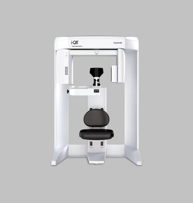
Your orthodontic treatment is only as good as the information behind it. A treatment plan built from a flat, two-dimensional X-ray and a visual exam is working with incomplete data. The i-CAT FLX Cone Beam 3D Imaging System changes that — completely.
In a single 8-second scan, Dr. Cosmo receives a full three-dimensional view of your teeth, roots, jaw, airway, and facial structure. That’s not just more detail — it’s a fundamentally different level of diagnostic accuracy that leads to more precise treatment planning, fewer surprises mid-treatment, and better outcomes overall.
Cranston Orthodontics was the first practice in Rhode Island to offer cone beam 3D imaging. It’s one of many ways we’ve consistently brought the most advanced tools in orthodontics to our patients before anyone else in the region.
0 Sec
Full Diagnostic Scan
First
In RI: Cone Beam 3DLower
Dose Than 2D X-Ray360°
Full Facial & Jaw ViewKey Features — i-CAT FLX Technology
One 8-Second Scan. A Complete Picture.
Traditional imaging captures flat, two-dimensional slices of your teeth and jaw. The i-CAT FLX captures a full three-dimensional image of your entire dental and facial structure in a single 8-second scan — giving Dr. Cosmo a level of diagnostic detail that 2D X-rays simply cannot provide. Less time in the chair. More information for your doctor.
01
Lower Radiation. Higher Clarity.
One of the most common patient concerns about imaging is radiation exposure. The i-CAT FLX’s QuickScan+ technology delivers full 3D dentition imaging at a radiation dose lower than a standard 2D panoramic X-ray. You get more diagnostic information at less exposure — a meaningful upgrade in both safety and precision.
02
Designed Around Patient Comfort
The i-CAT FLX was engineered with the patient experience in mind. The open-environment design eliminates the claustrophobic feel of traditional imaging equipment. The Ergonomic Stability System minimizes patient movement during the scan, reducing the need for retakes and keeping your appointment on schedule.
03
Smarter Diagnosis. Better Treatment Plans.
Visual iQuity™ advanced image technology delivers crisp, precise 3D and 2D images that allow Dr. Cosmo to evaluate bone structure, airway, tooth positioning, and soft tissue with a level of accuracy that changes what’s possible in treatment planning. Patients treated with i-CAT 3D imaging benefit from more targeted, more predictable outcomes.
04
The Right Scan for Your Specific Case
No two patients require the same imaging. SmartScan STUDIO™ allows Dr. Cosmo to select the exact scan type and field of view your case calls for — keeping radiation exposure As Low As Reasonably Achievable (ALARA) while capturing everything needed for a complete diagnosis.
05
What We Use 3D Imaging for
Application
Orthodontic Evaluation & Treatment Planning
TMJ Assessment
Airway & Sleep Apnea Screening
Impacted Tooth Evaluation
Full Facial & Jaw 3D Imaging
Bone & Soft Tissue Analysis
What It Helps Us Do for You
Creates a precise 3D model of your teeth, roots, and jaw to build the most accurate treatment plan possible.
Maps the temporomandibular joint in three dimensions to evaluate dysfunction, bite alignment, and joint health.
Identifies airway constrictions and structural factors that contribute to breathing difficulties and sleep-disordered breathing.
Locates and assesses impacted teeth with precision unavailable through traditional X-rays, guiding safer treatment decisions.
Captures the full craniofacial structure to support complex cases including jaw surgery evaluation and facial growth analysis.
Evaluates bone density and soft tissue structure to plan implants, extractions, or advanced orthodontic mechanics.
Why 3D Imaging Matters
The Difference Between a Good Treatment Plan and the Right One
When Dr. Cosmo evaluates your case using i-CAT 3D imaging, he’s not working from a shadow of your anatomy. He’s seeing it in full — every root, every millimeter of bone, every structural nuance that affects how your teeth will move and respond to treatment.
That matters because orthodontics isn’t just about aesthetics. It’s about occlusion, airway, joint health, and long-term stability. A 3D image lets Dr. Cosmo evaluate all of these factors together, at once, before a single bracket is placed or an aligner is designed.
The result is a treatment plan that accounts for who you are as a patient — not just what your teeth look like from the front.
Experience the Most Advanced Diagnostic Imaging in Rhode Island Orthodontics.
Schedule a complimentary consultation at Cranston Orthodontics and experience the difference that 3D precision makes from your very first appointment.