59 Phenix Ave Cranston, RI 02920
The X-Ray That Shows the Whole Picture
A standard dental X-ray provides a flat, two-dimensional view, revealing only part of the story. Important details like root positions, bone density, airway dimensions, jaw joints, and the spatial relationships between structures require a third dimension — which cone beam CT provides.
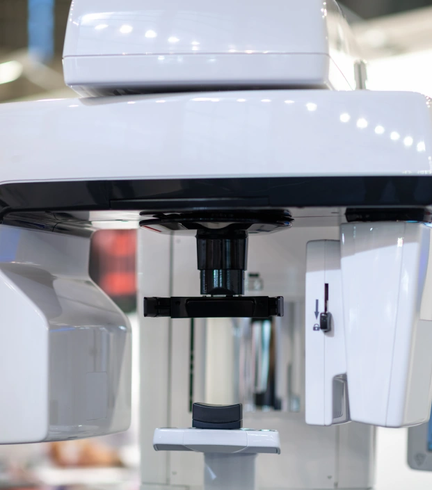
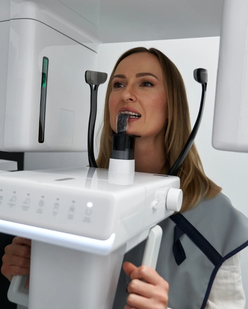
At Cranston Orthodontics, Dr. Cosmo uses cone beam CT as a core diagnostic tool, capturing a complete 3D image of each patient’s dental and skeletal anatomy in a fast, low-dose scan. This volumetric image can be rotated, sliced, and analyzed from any angle, delivering a level of clarity and precision that traditional radiography cannot match.
This matters because orthodontics is inherently three-dimensional — teeth move through bone, airways occupy space, and jaw joints function in all directions. Planning treatment with only 2D images is possible, but incomplete. Cone beam CT removes that limitation, enabling more accurate, fully informed treatment decisions.
0 Scan
Full craniofacial dataset
3D
Complete Volumetric ImagingLow Dose
Safe for Children and AdultsSeconds
Scan Acquisition TimeKey Features of Cone Beam CT Imaging
Complete 3D Craniofacial Data in One Scan
A single cone beam CT scan captures the teeth, roots, jaw, airway, temporomandibular joints, and surrounding bone in one continuous volumetric dataset. No repositioning, no separate scans for different regions, no missing information. Everything Dr. Cosmo needs to understand your anatomy in three dimensions is captured in a single acquisition that takes only seconds.
01
Airway Assessment — What Standard X-rays Cannot Show
The airway is invisible on conventional dental X-rays — but fully visible in three dimensions on cone beam CT. Dr. Cosmo uses airway imaging to assess the dimensions and geometry of the nasal and pharyngeal airway, identify areas of constriction, and evaluate the structural contribution of jaw position and arch width to airway patency. For children with breathing or sleep concerns, this data is clinically essential.
02
Root and Bone Visualization
Cone beam CT reveals the full three-dimensional position of every root — including roots that overlap or cross on conventional 2D X-rays and are therefore impossible to accurately localize without volumetric imaging. Bone density, root length, proximity to adjacent structures, and the available bone envelope for tooth movement are all visible and measurable — allowing Dr. Cosmo to plan tooth movements with a precision that 2D records cannot support.
03
Growth and Skeletal Assessment
For growing patients, cone beam CT provides a precise record of skeletal development — the three-dimensional relationship between the upper and lower jaw, the vertical dimension of facial growth, and the spatial position of developing permanent teeth within the bone. This data allows Dr. Cosmo to time orthodontic and growth-modification interventions with a level of precision that cephalometric X-rays alone cannot deliver.
04
Low Dose — Safe for Every Patient
Cone beam CT at Cranston Orthodontics uses a carefully calibrated low-dose protocol — delivering the comprehensive 3D diagnostic data required for thorough orthodontic assessment while keeping radiation exposure to the minimum clinically appropriate level. The scan is safe for children, teens, and adults, and is acquired in seconds with the patient seated comfortably upright — no lying down, no lengthy procedures.
05
How Cone Beam CT Connects to Your Full Treatment
Cone beam CT is the diagnostic foundation on which Dr. Cosmo builds every complex treatment plan at Cranston Orthodontics — connecting directly to every element of your care.
- Airway evaluation — 3D airway imaging data is used to assess airway dimensions and identify structural contributors to breathing concerns — informing referrals to ENT and sleep medicine specialists where indicated, and guiding orthodontic treatment decisions with direct airway implications.
- Growth analysis — Cone beam CT data provides a precise three-dimensional record of skeletal development — used to determine the optimal timing for early intervention, functional appliance therapy, and surgical planning in growing patients.
- Treatment planning via OrcheStrate 3D — Cone beam CT data feeds directly into OrcheStrate 3D, where Dr. Cosmo integrates it with iTero® digital scans, clinical photography, and cephalometric analysis to plan treatment in full 3D — visualizing tooth movements within the actual bone architecture of each patient's anatomy.
- Surgical planning — For patients requiring corrective jaw surgery, cone beam CT provides the three-dimensional skeletal data required for precise virtual surgical planning — including the exact repositioning of the jaws and the anticipated changes to the facial profile and airway.
- Impacted teeth — Cone beam CT reveals the precise three-dimensional location of impacted or unerupted teeth — including their relationship to adjacent roots, nerve canals, and the overlying bone — allowing Dr. Cosmo to plan exposure and eruption guidance with confidence.
- iTero® integration — Cone beam CT data and iTero® digital surface scans are combined within the diagnostic workflow to create a complete picture of each patient's anatomy — surface detail from the iTero® scan, internal bone and airway data from cone beam CT — giving Dr. Cosmo the most comprehensive diagnostic dataset available in modern orthodontics.
When You Can See Everything, You Can Treat Everything — With Confidence.
At Cranston Orthodontics, cone beam CT is not reserved for complicated cases or surgical referrals. It is part of how Dr. Cosmo approaches every patient who requires comprehensive diagnostic assessment — because the most accurate treatment decisions begin with the most complete picture of the anatomy. For children with airway or growth concerns, for adults with complex bite and jaw issues, and for any patient whose care benefits from knowing exactly what lies beneath the surface, cone beam CT is the difference between treating what you can see and treating what's actually there.
Schedule a Free Consult